Wednesday, May 8, 2013

Stem Cell Transplantation Touching Lives


Our bustling lives are an epitome of the human splendor as well as the challenges we have instituted for ourselves. One such challenge is being healthy! Good health is undoubtedly the germ of optimism, which is rarely found in the modern lifestyle. While we have managed to run ahead of our needs, even to the extent of surpassing our wants, the newfound lifestyle has brought a decline in the overall health and well-being of our society. Factors like imbalanced diet, junk food, exposure to toxic content, polluted atmosphere are to name a few, which have led to lethal diseases like cancer having devastating effects on one's body in a long run, if not instant to be propitious. Thankfully, our modern healthcare facilities have not disappointed us at all. Institutes like Apollo hospitals and similar research and healthcare organizations have left no stone unturned to come up with modern healthcare facilities touching and soothing millions of lives daily.


We have seen times when a number of hematological disorders like leukemia (cancer of blood), beta thalassemia (genetic blood disorder), severe aplastic anemia (bone marrow does not produce sufficient new cells to replenish blood cells) etc. were invariably fatal, and there was no cure for them. Fortunately, our modern and well-equipped medical system is advancing with a great pace, and one such development is the use of "stem cell transplantation" technique which has arisen hopes for fighting such acute medical conditions.

Stem cell transplantation is an upcoming and a revered field in modern medicine. It involves the infusion or injection of healthy stem cells into the body to replace the damaged or diseased cells. Stem cell transplantation can be of three types depending from where cells have been used; as autologous transplantation (cells from your own body), allogenic stem cell transplantation (cells from a donor) or syngenic transplantation (cells from identical twin). Also, the stem cell transplantation is a general term and it can be called specifically by different names such as bone marrow transplant, peripheral blood stem cell transplant or an umbilical cord blood transplant depending on the source of the stem cells. Traditionally, donated organs or tissues were substituted for damaged or dysfunctional ones. However, the sad part of this story was that number of people waiting for transplantation outnumbered the number of available donors. As a result, people died awaiting transplants. Here, stem cells have a great potential to offer as a source of replacement to treat various diseases and reduce the morbidity and mortality of such awaiting patients.

The stem cells have many current applications for treating various diseases which include severe burns, type I diabetes, aplastic anemia, and its potential to treat many more diseases in the future such as neurological disorders (including Parkinson's and Alzheimer's) is being researched upon and surely it will pave the way for treating many other diseases. According to Lalit Kumar, professor of medical oncology at AIIMS, stem cell transplants have shown an around 50% of success in treating certain kinds of cancer and even a higher around 70 – 80% of success in treating other diseases like beta thalassemia, aplastic anemia.

India has a few centers/hospitals where stem cell transplantation can be done as per the needs. One such prestigious center is Apollo hospitals (http://www.apollohospitals.com/cutting-edge.php). This hospital (Dr. Chirag Shah, Apollo International hospitals) is known to have successfully done the first autologous stem cell transplant for acute myeloid leukemia, and first allogenic stem cell transplant for leukemia in private sector of Gujarat.

Today, stem cell transplantation do offer hope for effective treatment with infinite possibilities in modern day healthcare. Moreover, stem cells can be used for research purposes for screening new drugs before being used on humans. They can even act as a testing model without risking the health of the volunteer. The continued research will make a way for the more incurable diseases and disorders, and time will witness more and more success stories of stem cell therapies, touching and reassuring more lives.


Top Image Source: http://www.apollohospitals.com/stem-cell-transplant.php
 

Thursday, April 25, 2013

Biomolecules of the Cell - Nucleic Acids (Part 2)

In the previous post, we have seen the basics of nucleic acids and the native form of DNA i.e.; B-DNA in details.
There are various other forms which DNA can assume. In this post, I will discuss some of the other forms of DNA (i.e.; A-DNA and Z-DNA) and another type of nucleic acid - RNA.
Starting the post with the A-form of DNA.

A-form of DNA: 
The following are certain characteristics of A-DNA:
i. This form of DNA exists when the relative humidity is around 75%.  
ii. The A-DNA is also right-handed helix like B-DNA.
iii. A-DNA is shorter and wider than B-DNA. 
iv. The diameter of A-DNA is 23Å as against 20Å in B-DNA. 
v. There are 11 base pairs per turn (more than B-DNA which has 10 base pairs per turn). 
vi. The rise per turn is 28Å. 
vii. Thus, if we calculate, rise per base pair (28Å/11base pairs), we get 2.6Å. 
viii. The base pairs are not perpendicular to the helical axis instead they are tilted 20˚ w.r.t. helical axis. 
ix. Just like B-DNA, A-DNA also has major groove and minor groove; however, the former is very deep and latter is very shallow as can be seen in the adjacent diagram.

Does A-DNA have any biological importance? Yes. A-DNA is observed in some biological context as follows. 
A ̴ 3 base pair segment of A-DNA is present at the active site of DNA polymerase.
Also, gram positive bacteria undergoing sporulation contain a high proportion (20%) of small acid soluble-spore proteins (SASPs). Some of these SASPs induce B-DNA to assume the A-form of DNA (atleast in vitro).
Another instance where A-form of DNA is present is during RNA-DNA hybrids. RNA-DNA hybrids do occur in the cell at different junctions for example, during replication (where replication is initiated by RNA primer) and also during transcription (where RNA is made on DNA template). These RNA-DNA hybrids cannot take up the B-form of DNA (because of the 2' oxygen of RNA) and hence, they resemble A-form of DNA.

Z-form of DNA:
Another form of DNA is Z-DNA and is called so because of its zigzag pattern of the phosphate backbone. The following are some of the characteristics:
i. The main characteristic is that Z-DNA is a left-handed double helix.
ii. The formation of Z-DNA is base sequence dependent (composition dependent). Only alternating purine and pyrimidine polymers can form Z-DNA. Thus, this zigzag forms the repeating unit of Z-DNA which is a dinucleotide. 
iii. The right handed DNA can be transited to left handed DNA (Z-DNA) in solutions that include high ionic strength, hydrophobic solvents, presence of certain trivalent cations or covalent modification with bulky alkylating agents.
iv. It contains approximately 12 base pairs per turn of the helix. 
v. Rise per turn of the helix is 44Å.
vi. The helix rises by 2.7Å per base pair. 
vii. The base pairs are perpendicular of the helix (just like that of B-DNA).
viii. Regarding the grooves in Z-DNA, it possesses only a minor groove. There is no discernible major groove and the minor groove is extremely deep and narrow.

The following table will give you an idea of major structural differences between three major forms of DNA (B-DNA, A-DNA and Z-DNA).


Ribonucleic Acid (RNA):
Coming to another type of nucleic acid which is RNA. As we have seen the major difference between RNA and DNA in previous post, here I will discuss about its types and structure. 
Types of RNA:
There are various kinds of RNAs. Here, the RNAs have been classified into two major groups as coding RNAs and non-coding RNAs. 
Coding RNA - mRNA
a. Coding RNAs:
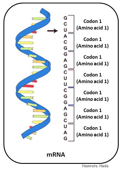
mRNA (messenger RNA) is coding RNA that is involved in the process of translation in the cell. It is called messenger as it carries information from DNA to the ribosome which is the site of protein synthesis. The coding sequence of mRNA determines the amino acid sequence in the protein that is produced.

b. Non-coding RNAs:
Most of the RNAs do not code for any proteins and these types of RNA fall under another group as non-coding RNAs (ncRNAs). These so called non-coding RNAs can be encoded by their own genes. The most prominent examples are rRNA (ribosomal RNA) and tRNA (transfer RNA), both of which are involved in the process of translation.
Some other examples of noncoding RNAs are those involved in gene regulation and RNA processing. There are also some non coding RNAs that are able to catalyse chemical reactions such as cutting and ligating other RNA molecules and catalysis of peptide bond formation in ribosome. Such catalytic RNAs are called ribozymes. 

Structure of RNA:
The structure of RNA is described as follows:
Primary Structure of RNA:
Primary structure of RNA is somewhat similar to that of DNA; the only difference being the sugar (ribose) component which has an additional hydroxyl group at 2' carbon and thymine is replaced with uracil (see figure on the left).
Note: The hydroxyl group on the 2' carbon atom of ribose make it more labile as compared to DNA and it provides a chemically reactive group. Because of this lability, RNA is cleaved into mononucleotides by alkaline solution and DNA is not.
RNA is a polynucleotide chain that can be double stranded or single stranded, linear or cicular. 

Secondary Structure of RNA: 
There are various types of RNA exhibiting different conformations. The simplest secondary structures in single stranded RNAs are formed by pairing of complementary bases. Two major forms of secondary structures are hairpins and stem loops.
‘Hairpins’ are formed by pairing within approximately 5-10 nucleotides of each other whereas in stem loop, there is pairing of bases that are separated by approximately 50 to several hundreds of nucleotides.

Tertiary Structure of RNA:
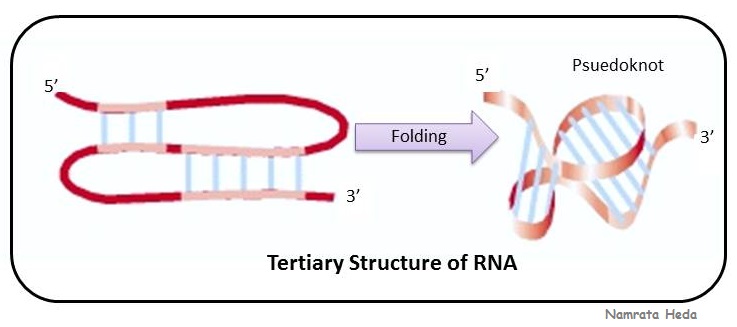
These simple folds (hairpin and stem-loops) can cooperate to form more complicated tertiary structures, and one of such structure is 'pseudoknot' as can be seen in the adjacent figure.
tRNA is a well-defined example of tertiary structure of RNA. tRNA is a type of RNA that is involved in translation process of protein synthesis. They have a L-shaped 3D structure that allows them to fit into the P and A-sites of the ribosome. The figure depicted as tRNA above under non-coding RNAs is the three-dimensional structure of tRNA.

Here, we complete the nucleic acids topic. Any comments or doubts are welcome.!

Friday, April 19, 2013

Biomolecules of the Cell - Nucleic Acids (Part 1)

Nucleic acids are considered to be the building blocks of all the living organisms. The building blocks of nucleic acids are nucleotides. DNA (deoxyribonucleic acid) and RNA (ribonucleic acid) are the nucleic acids. They can be described as the polymers of nucleotides linked through phosphodiester bonds. Don't worry, before going ahead and learning more about nucleic acids, we will go through the basics. Keeping in mind the basics, lets will start with nucleotides.

Bases, Nucleosides and Nucleotides:
Nucleotide has a distinctive structure and is composed of the following components which are bound together covalently:
a. Base (contains nitrogen) – This can be either a pyrimidine or a purine (explained below)
b. Sugar (5-carbon or pentose) – This can be either ribose or deoxyribose
c. A phosphate group
When a base and sugar is present (no phosphate), then it is called a nucleoside as can be seen in the adjacent figure.
When all the three components (base, sugar and phosphate) are bonded together, then this is known as nucleotide. Nucleotides can also exist in activated forms containing either two phosphates (diphosphate) or three phosphates (triphosphate).
When the sugar in the nucleotide is ribose, then the nucleotide is called ribonucleotide and when the sugar is deoxyribose, then nucleotide becomes deoxynucleotide.
Let us have a look at the structure of a nucleotide and make the concepts all the more clear. In this adjacent figure, the structure of deoxyadenosine monophosphate will depict all the three components as sugar, base (here, deoxyribose) and phosphate. In comparison, the diagram on the right has an extra hydroxyl group (-OH) on 2’ carbon atom of ribose sugar, making it ribonucleotide (instead of deoxyribonucleotide).  Also, in this diagram, note the 5’ and 3’ carbon atoms. If we understand these 5' and 3' carbon atoms, this will aid in understanding the polarity of the nucleic acids. The 5’ carbon atom is attached to the phosphate group while 3’ carbon atom is attached to a hydroxyl (-OH) group. 

There are five bases known as Adenine (A), Guanine (G), Cytosine (C), Uracil (U) and Thymine (T). The point to remember is that Uracil is not present in DNA but present in RNA while Thymine is not present in RNA, but present in DNA. Here is the table showing all the five bases with their structure, abbreviations and their nucleoside and nucleotide forms.
Another way to categorize nucleotide bases is as ‘purines’ and ‘pyrimidines’. Purines include A and G (which are double-ring members) while pyrimidines include the remaining T, C and U (which are single-ring members). A point to remember is that in double-stranded nucleic acids, the pairing is always between a purine and a pyrimidine.


Polynucleotides:
The polynucleotides are polymeric compounds consisting of 15 or more nucleotide monomers covalently bonded together in a chain. As we have seen above, the carbon atom in the sugar is numbered 1’ to 5’. The hydroxyl (–OH) group on the 3’ carbon of one nucleotide can react with the phosphate attached to the 5’ carbon of another (adjacent) nucleotide to from a dinucleotide held together by phosphate ester bonds. These bonds are also called phosphodiester bonds. As the chain contributes more and more nucleotides to itself; it begins to grow and hence, becomes a polynucleotide.
A short segment in the adjacent figure will make it all the more clear. Remember that DNA is read from 5’ end to the 3’ end. If we read this DNA segment, then the sequence goes like this – adenine (A), cytosine (C), guanine (G) and thymine (T).

DNA and RNA are the main polynucleotides. First, we will have a look at DNA.

DNA (Deoxyribonucleic Acid):
DNA is the molecule that encodes the blueprint of an organism meaning the DNA contains all the information required to build up and maintain an organism. Let me ask you one question, by whom was DNA first discovered? You might be thinking, Watson and Crick..!! Let me make it clear to you, the answer here is NO. The DNA was first discovered in 1868 by Swiss-physician Friedrich Miescher. He isolated a compound from the nuclei of white blood cells. This compound was neither a protein, nor a carbohydrate, nor a lipid but a unique type of biomolecule. Miescher named it ‘nuclein’ as he had isolated it from the nuclei of the cell. Today, this molecule is called DNA.


Erwin Chargaff was a biochemist who did an extensive research on chemical analysis of the base composition of DNA and stated that the base composition of DNA varied from organism to organism but was independent of age, sex, nutritional status or any other environmental factors. He came up with certain relationships which were called the 'Chargaff’s rule' and they are as follows:
a. The amount of adenine is always equal to the amount of thymine (A=T).
b. The amount of guanine is always equal to the amount of cytosine (G=C).
c. The amount of A+G is 50% of the total amount of bases in the molecule.
d. The amount of T+C is 50% of the total amount of bases in the molecule.


 Then, what was the contribution of Watson and Crick. So, here lies the answer - in 1953, James D. Watson and Francis Crick described the molecular structure and shape of DNA.
When the counterion is Na+ and the relative humidity is 92%, then DNA fibers assume what is known as “B conformation”. I would like to jot some points/features of B form of DNA from my HOD, Dr. Avinash Upadhyay’s book “Molecular Biology” because they are so beautifully explained in the book that I still remember them even after years. 
Watson and Crick described the shape of DNA as ‘double helix’ and following are the features of B-DNA:
i. Double-stranded helix: DNA consists of two polynucleotide strands coiled around each other to form a double stranded helix.
ii. Plectonomic coil: The two strands are coiled around each other in such a way that they cannot be separated without unwinding the helix (plectonomic coil).
iii. Antiparallel: The two strands are antiparallel to one another meaning they run in opposite directions. Both the strands have one 5' phosphate terminus and one 3’ hydroxyl (-OH) terminus. Antiparallel strands mean that the 5’ terminus of one strand is adjacent to 3’ adjacent of another strand and vice-versa (as can be seen in the adjacent figure).
iv. Right handed helix: The helix is a right-handed helix.
v. Diameter: The diameter of the helix is 20Å.
vi. Sugar phosphate backbone - Hydrophilic: The sugar-phosphate backbones of both the strands are hydrophilic in nature and follow the helical path and are towards the outer edge of the molecule where they will be able to interact with the aqueous environment.
vii. Bases - Hydrophobic: The bases are hydrophobic in nature and they are placed at the interior of the helix.
viii. Planar base pairs: The planes of the bases are perpendicular to helical axis. Each base is hydrogen bonded to a base on the opposite strand to from a planar base pair. Two types of purine-pyrimidine base pairs can occur as A..T and C...G. There are two hydrogen bonds between A and T and three hydrogen bonds between C and G. This is the reason why the helix has a constant diameter of 20Å.
xi. Complementary base pairing: This specificity of the base pairing is referred to as ‘complementary base pairing’.
x. Thickness: The bases have a Vander Waal’s thickness of 3.4Å.
xi. Base pairs per turn: There are 10 base pairs per turn of the helix and hence, the helix rises by 34Å per turn.
xii. Rotation per base pair: If there are 10 base pairs per turn, then each base pair rotates by 36˚ (as one rotation is 360˚ and divided by 10 will give 36˚) and hence, a 36˚ turn per base pair is present.
xiii. Major and minor grooves: The angle of the glycosidic linkages lead to the formation of two external helical grooves. One of the groove is deep and wide and is called the major groove. Another groove is shallow and narrow. This is the minor groove.

This was all about B-form of DNA. There are some other forms of DNA as well, which we will discuss in the next post along with RNA.